By Katie Kozlowski
The great saphenous vein (GSV) is a large, superficial vein arising from the dorsal pedal venous arch on the dorsal (top) aspect of the foot. It runs medially along the inside of the leg and thigh to join the common femoral vein 3-4 cm below the public bone. (Meissner, 2005)
The GSV lies in the saphenous compartment, a superficial compartment in the leg bordered by muscular fascia deeply and the hyperechoic saphenous fascia superficially. In the thigh, the GSV can be readily visualized in saphenous compartment under ultrasound guidance. The appearance of the GSV and saphenous compartment under ultrasound is often referred to as the “Egyptian Eye”. See image below. (Cavezzi et al, 2006)
Venous insufficiency in the great saphenous vein can be treated with endovenous laser ablation, radiofrequency ablation, VenaSeal (glue), MOVA, or ultrasound guided foam sclerotherapy.
(Meissner, MH. Lower Extremity Venous Anatomy. Semin Intervent Radiol 2005; 22(3): 147-156.)
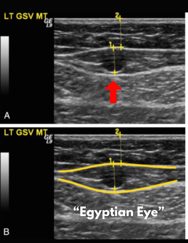
FIG. 1
(A) Transverse B-mode image of the great saphenous vein (
GSV ) with measurements of vein diameter (
1 ) and depth from surface (
2 ). (B) The vein is surrounded by the echogenic superficial (
top outline ) and muscular fascia (
bottom outline ) forming the saphenous eye, Egyptian eye, or eye of Horus.
(Modified from Cavezzi A, et al: Duplex ultrasound investigation of the veins in chronic venous disease of the lower limbs—UIP consensus document. Part II. Anatomy.
Eur J Vasc Endovasc Surg. 2006;31[3]:288–299.)